

Researches
This page summrizes the researches I have been working on. Which can be roughly divided into the following categories:
-
Fourier ptychographic imaging
-
Lensless imaging
Fourier ptychographic imaging
Fourier ptychographic (FP) imaging is a recently developed technique for wide field, high resolution, and quantitative phase imaging. FP applies the concepts from ptychography, synthetic aperture and phase retrieval.
In a typical FP system, a programmable light emitting diode (LED) array serves as a partially coherent light source to provide angularly varying oblique plane wave illuminations. At each illumination angle, the image sensor records a low-resolution intensity image of the specimen. The captured low-resolution images contain different information in the Fourier domain and can be synthesized to recover a high-resolution image.
We build a Fourier ptychographic microscopy (FPM) experimental system by adding a LED illumination module to a commercial microscopy and make several modifications based on the system.

The setup pf our FPM experimental system.

The reconstruction procedure of FPM.
We proposed a modified global reconstruction method for FPM termed gradational FPM (gFPM), which significantly improves the convergence rate and reconstruction result. [1]

Intensity and phase SSIM curves varying with noise standard deviation and the reconstruction results of three methods with 7 × 10⁻⁴ noise standard deviation.
We propose a preprocessing LED array alignment method for FPM termed BFL (brightfield localization) for FPM which corrects the global misalignment of LED by utilizing the spatial relationship between the LED elements and the brightfield locations on the sample plane. [2]

The flow diagram of applying BFL in FPM reconstruction. The BFL includes three steps that are preprocessing, PSO method and RANSAC method.
We propose an efficient colorful FPM reconstruction method using multi-resolution wavelet color fusion which achieves better result and speed compared with conventional methods. [3]
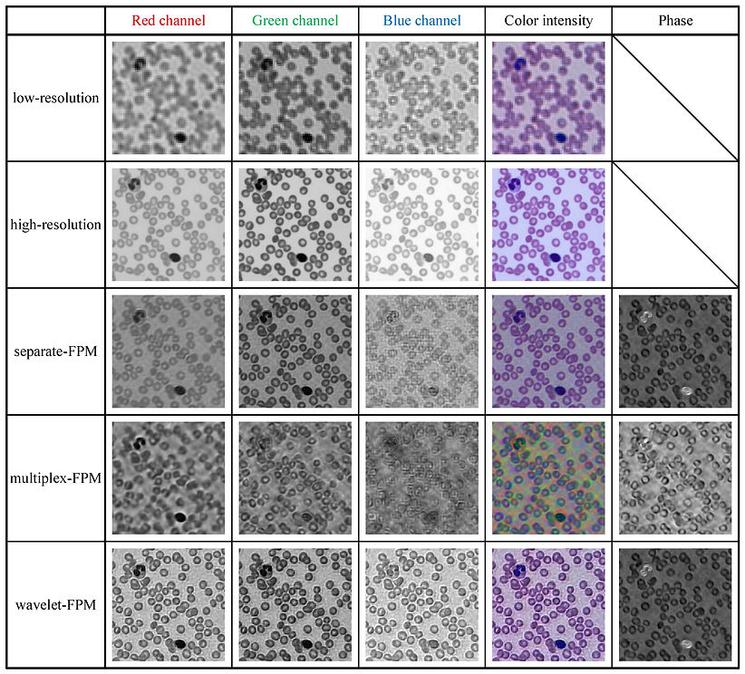
Reconstruction performance of three methods on human blood smear images.
-
Jizhou Zhang, J. Z., Tingfa Xu, T. X., Xing Wang, X. W., Sining Chen, S. C., & Guoqiang Ni, G. N. (2017). Fast gradational reconstruction for Fourier ptychographic microscopy. Chinese Optics Letters, 15(11), 111702. https://doi.org/10.3788/COL201715.111702. [Download PDF]
-
Zhang, J., Xu, T., Liu, J., Chen, S., & Wang, X. (2018). Precise Brightfield Localization Alignment for Fourier Ptychographic Microscopy. IEEE Photonics Journal, 10(1), 1–13. https://doi.org/10.1109/JPHOT.2017.2780189. [Download PDF]
-
Zhang, J., Xu, T., Chen, S., & Wang, X. (2018). Efficient colorful Fourier ptychographic microscopy reconstruction with wavelet fusion. IEEE Access. https://doi.org/10.1109/ACCESS.2018.2841854. [Download PDF]
Lensless imaging
High-resolution optical microscopy traditionally relies on high-magnification and high–numerical aperture objective lenses. Lensless microscopy can provide high-resolution images without the use of any focusing lenses, offering the advantages of a large field of view, high resolution, cost-effectiveness, portability, and depth-resolved three-dimensional (3D) imaging.
We are now building a lensless microscopy system and trying to reconstruct images using the transport of intensity equation (TIE).
